
Causes of Parkinson's disease.
- Parkinson’s disease can be defined clinically as:
'A disorder of unknown cause which is characterized by a tremor that is present at rest, bradykinesia, stiffness of muscles and impairment of postural reflexes which can therefore lead to falls and injury. With this a patient may also suffer from some cognitive abnormalities ranging from subtle frontal lobe defects to severe dementia.'
- The disease can also be pathologically defined as a presence of Lewy bodies, with a loss of dopamine in the brain.

- Idiopathic Parkinson’s disease is known to be due to selective degeneration of dopaminergic neurons of the substantia nigra and other brainstem nuclei, but the cause of this degeneration not clearly understood. The surviving neurons contain Lewy bodies. It is the presence of Lewy bodies which is the hallmark of the disease.
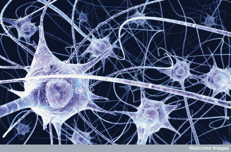
Dopamine
- A phenethylamine which functions as a neurotransmitter in the brain. It is also classified as both a catecholamine and a monoamine and is also a precursor of adrenaline.
- Dopamine is synthesized by the hydroxylation of L-tyrosine to L-DOPA using the enzyme tyrosine 3-monooxygenase or tyrosine hydroxylase. This product is then decarboxylated by dopa decarboxylase to form dopamine.
Image showing the biosynthesis of Dopamine courtesy of user NEUROtiker of Wikimedia Commons under the GNU free documentaion licence.
- Dopamine is stored in vesicles at nerve terminals and released upon the arrival of a presynaptic action potential down that nerve fibre. Dopamine can be broken down or will bind to specific dopamine receptors. In Parkinson’s disease the receptors are still available but the loss of dopamine producing nerves means there is a lack of dopamine to activate these receptors.
- Most of the dopaminergic nerve cells are found deep within the midbrain. From this area, connecting fibers release dopamine in many parts of the brain including the outer layer of the frontal cortex which is known to be involved in the limbic system.
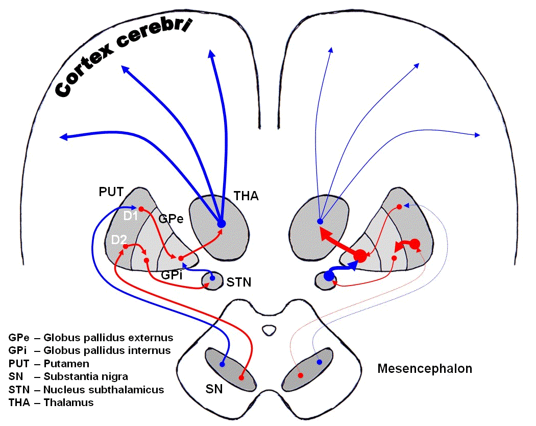
Basal Ganglia
- A group of structures found deep within the cerebral hemispheres and are thought to be involved mainly in movement control. They receive dopamine signals from the substantia nigra in the midbrain.
- The basal ganglia consist of a number of nuclei including the caudate nucleus, putamen, globus pallidus, subthalamic nuclei and the substantia nigra. Together the putamen and the caudate nucleus form the striatum, the location in which abnormalities can lead to movement dysfunctions including parkinsonism and ballismus. These dyskinesias can be seen in parkinson’s patients following drug treatment.
- The substantia nigra produces dopamine and sends connecting fibers or axons to the striatum. Here these fibers release dopamine allowing normal movement.
Nigrostriatal pathway
- Links the substantia nigra and the striatum.
- This pathway degenerates in Parkinson’s disease resulting in this loss of dopamine levels therefore leading to the characteristic symptoms of the disease. Other areas will also be seen to degenerate in Parkinson’s disease due to other parts of the nervous system being affected by loss of dopamine. For example, changes in the outer grey matter of the brain may be observed which are thought to be linked with some of the cognitive difficulties experienced in Parkinson’s disease.
Image showing dopaminergic pathways of the brain courtesy of user Chris 73 of Wikimedia Commons under the GNU free documentation licence.
It is in all sites of dopaminergic neuron cell loss or where the nerve cell is dying, Lewy bodies can be seen. Therefore Lewy bodies are most commonly seen in the substantia nigra. Lewy bodies are eosinophilic cytoplasmic inclusions consisting of a dense core primarily made up of alpha-synuclein.
Substantia Nigra histological sample image courtesy of user Filip em of Wikimedia Commons under the creative commons attribution licence.
